After three days, I weigh my samples again and the difference in their weight (before and after being three days in the extractor) reflects how much water was in the soil (remember peatlands are very wet?).
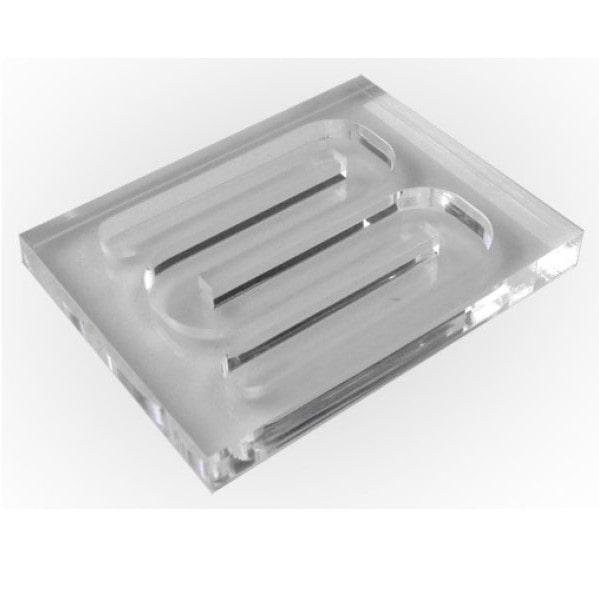
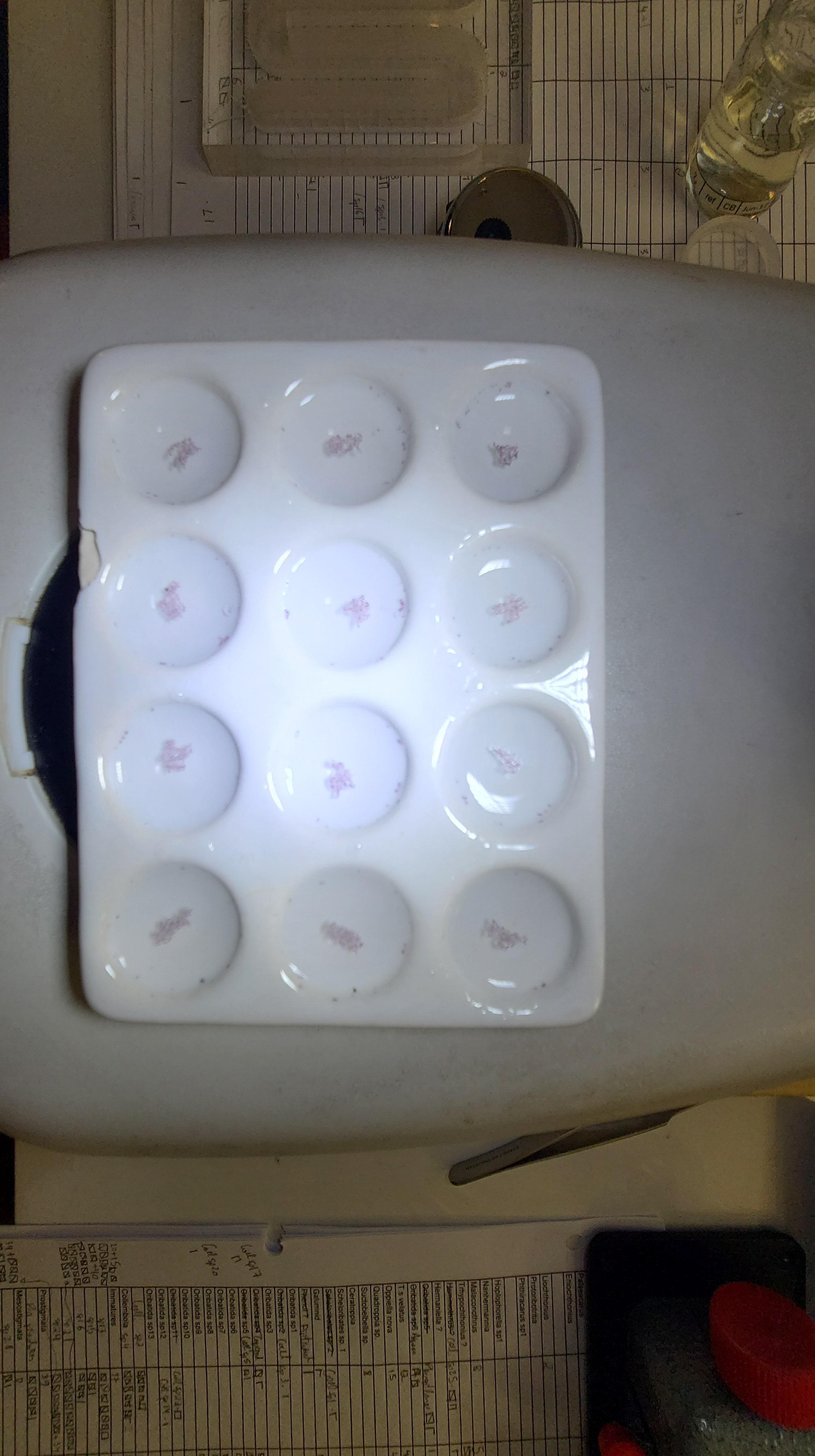
After these three days, I get my samples and pour them into a sorting dish called a "Bogorov chamber" (see picture). I look at the animals present in the ethanol through a microscope that can magnify up to 75x zoom! That’s already enough power to see most animals with clarity. Under this microscope, I count all the animals I see and separate them into major groups like beetles, oribatid mites, ants and so on.
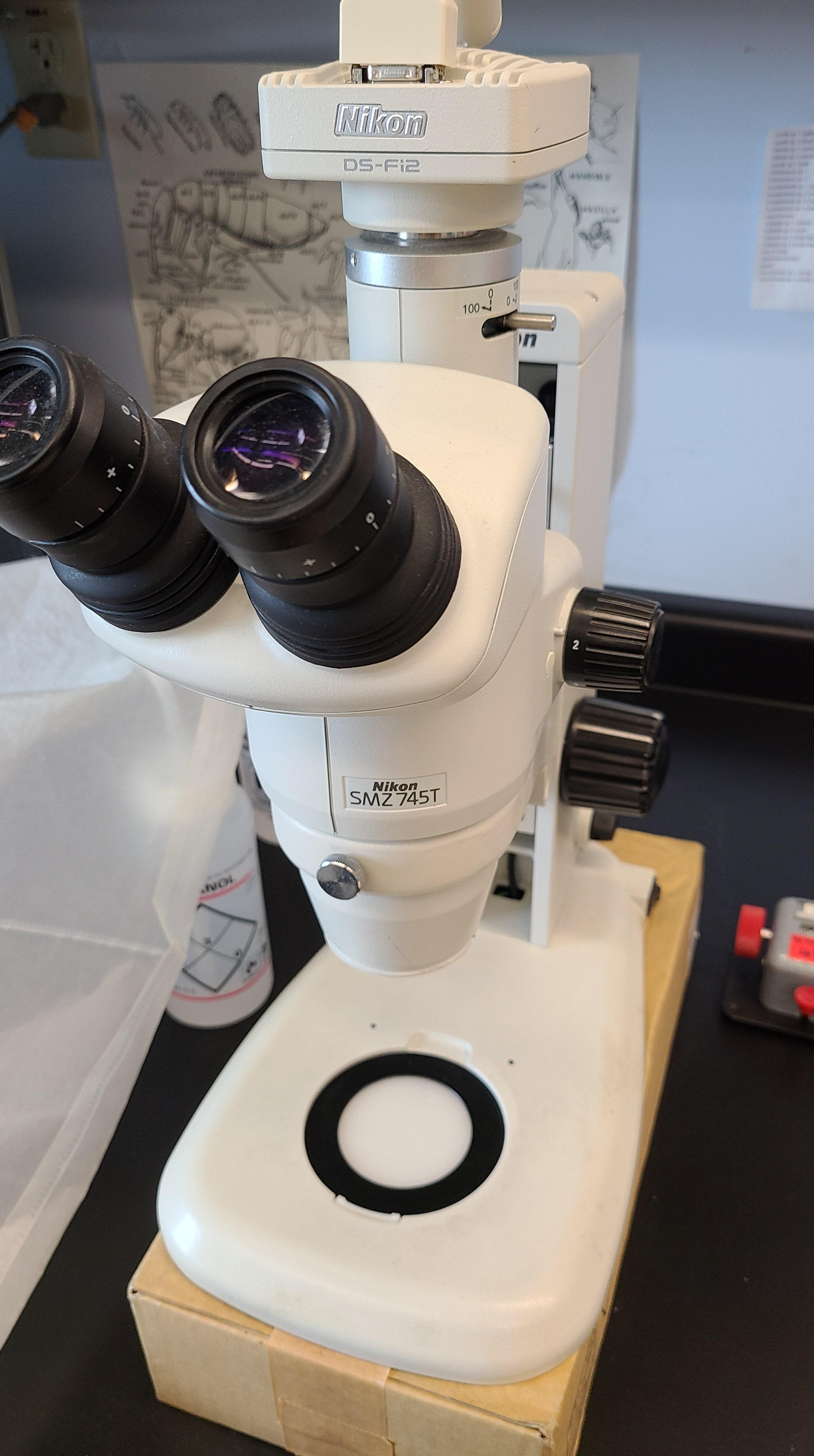
However, some oribatid mites are extremely small, so I need to look at them even closer. For those, I need to use another more potent microscope that augments in 400x what I see. This also helps to confirm the species identity of all oribatid mites. It’s incredible how many details this second (400x) microscope reveals, but it’s no quick task.
To use this second microscope, I need to prepare the individuals. This means getting an individual of oribatid mite of interest and putting it on a microscope slide. I use a chemical (not safe for children to use) to help highlight structures of its body under the light of the microscope. I can also use the camera attached to the microscope to take pictures. Did you see my first video call? There are lots of pictures of oribatid mites there too!
Since my focus is on oribatid mites, I only put them on slides to look closer and identify them. Other people in my lab work with other types of mites and their tasks are quite similar to mine. They use both microscopes too.