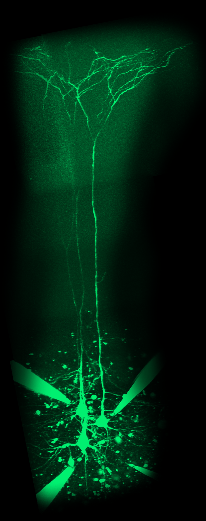
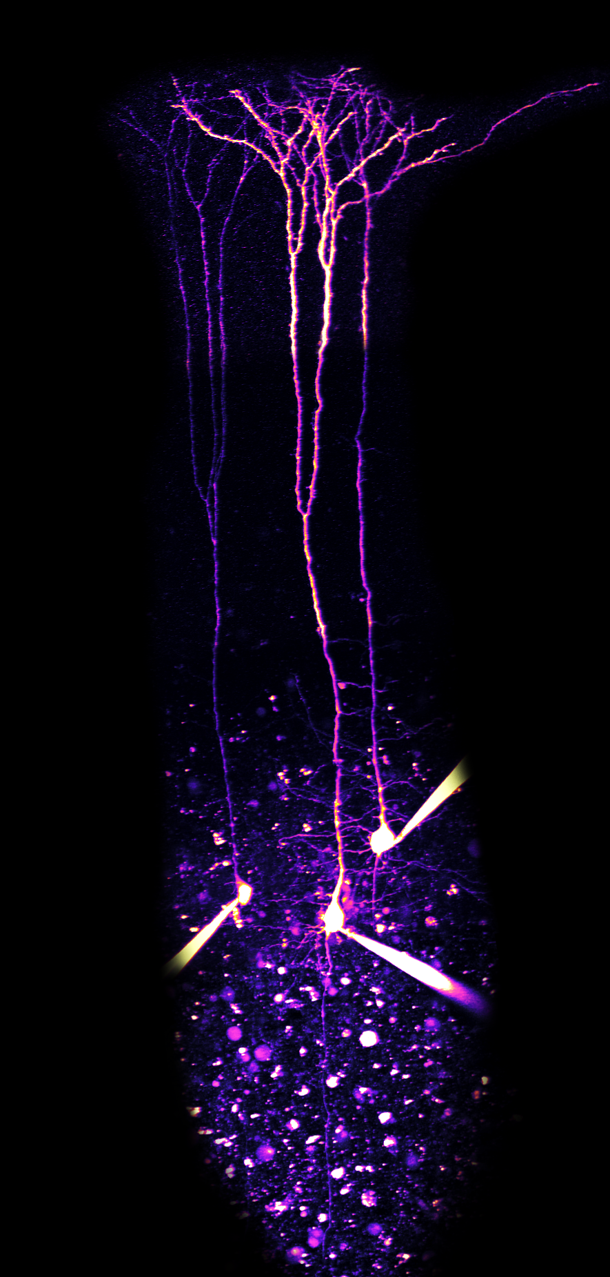
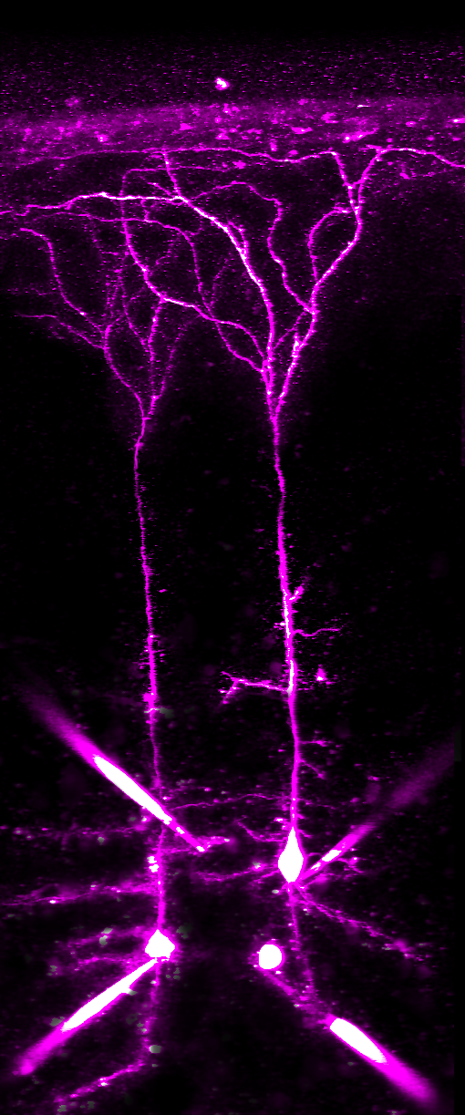
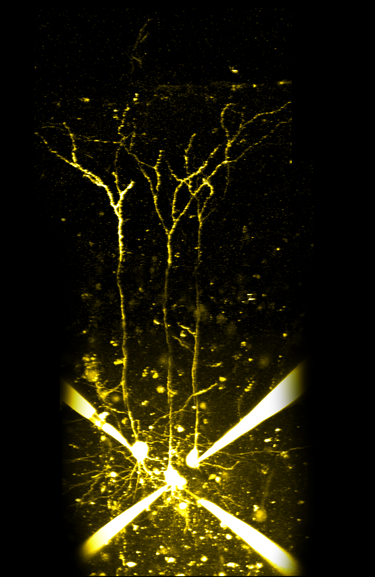
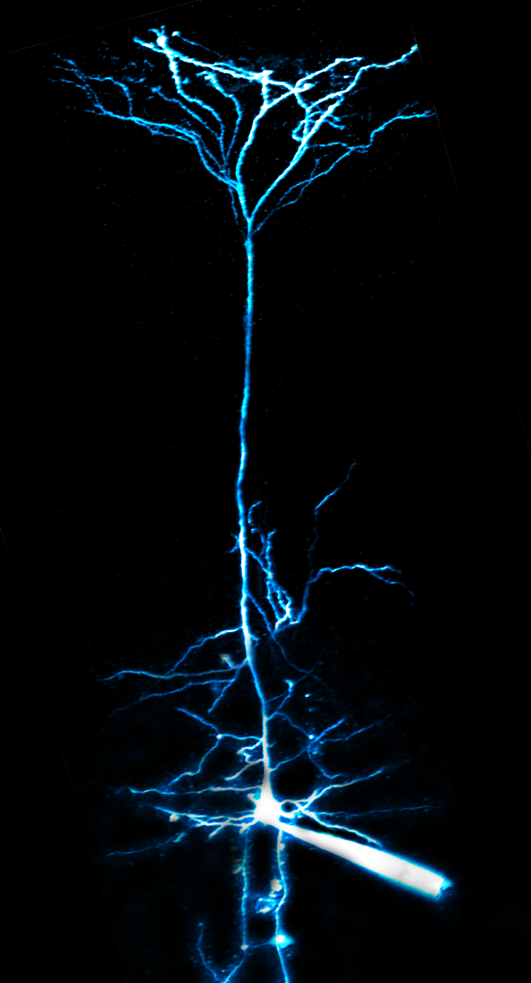
Imaging Neurons!

Photos

These thick tufted pyramidal neurons are my main target for my current study, and here is my most recent image!
In all the images, you can notice a bright line/thin triangle coming into frame on to or around the neuron being imaged... this is my patch clamp pipette!
The patch clamp pipette is filled with a fluorescent dye that is injected into the neuron when I "patch" it
You'll notice that this neuron is a bit different compared to the other ones, because it is most likely a basket cell, not a pyramidal cell... this image perplexed my peers and me as it appears the neuron is interacting with the blood vessel!
Basket cells are a type of interneuron, specifically inhibitory neurons that act through GABA... here are more four putative basket cells!
When I prep the neurons and prepare their tissue, sometimes I accidentally cut off the dendrites, which is bad for a lot of experiments, since neurons need their dendrites for their normal means of communication!
All the previous examples of pyramidal neurons have their spines and branches, but here's another example of pyramidal cells with all the lovely branches and spines!
Last but not least, here is my first cell I imaged in this lab; you'll see there is just one, because I had not yet learned how to patch multiple cells at once!

Videos